Alzheimer's Disease

What Is Alzheimer's Disease? What Causes Alzheimer's Disease?


Alzheimer's disease
is a progressive neurologic disease of the brain leading to the irreversible loss of neurons and the loss of intellectual abilities, including memory and reasoning, which become severe enough to impede social or occupational functioning. Alzheimer's disease is also known as simply Alzheimer's, and Senile Dementia of the Alzheimer Type (SDAT) .

During the course of the disease plaques and tangles develop within the structure of the brain. This causes brain cells to die. Patients with Alzheimer's also have a deficiency in the levels of some vital brain chemicals which are involved with the transmission of messages in the brain - neurotransmitters.

Alzheimer's disease is the most common form of dementia. The disease gets worse as it develops - it is a progressive disease. There is no current cure for Alzheimer's, although there are ways of slowing down its advance and helping patients with some of the symptoms. Alzheimer's is also a terminal disease - it is incurable and causes death.

According the National Institute on Aging, there are estimated to be between 2.4 million and 4.5 million Americans who have Alzheimer's. There are approximately 417,000 people in the UK with Alzheimer's, according to the Alzheimer's Society.

Why the name Alzheimer's disease? Aloysius Alzheimer and Emil Kraepelin, Germany

Aloysius Alzheimer was a German neuropathologist and psychiatrist. He is credited with identifying the first published case of "presenile dementia" in 1906, which Kraepelin later identified as Alzheimer's disease - naming it after his colleague.

In 1901, while he worked at the city mental asylum in Frankfurt am Main, Germany, Dr. Alzheimer had a 51 year old patient called Mrs. Auguste Deter. The patient had distinct behavioral symptoms which did not fit any existing diagnoses - she had rapidly failing memory, disorientation, confusion, had trouble expressing her thoughts, and was suspicious about her family members and the hospital staff. Her symptoms progressed relentlessly. Dr. Alzheimer wrote that she once said to him "I have lost myself."

Over the coming years Auguste Deter would take up more and more of Dr. Alzheimer's time, to the point of almost becoming an obsession for him. The lady died in 1906 and Dr. Alzheimer, who was working at Kraepelin's lab in Munich, had her patient records and brain sent there.

Along with two Italian doctors, Dr. Alzheimer performed an autopsy. The autopsy revealed that her brain had shrunken dramatically, but there was no evidence of atherosclerosis (thickening and hardening of the walls of the arteries). He used a silver staining technique he had learnt from ex-colleague Franz Nissl which identified amyloid plaques and neurofribrillary tangles in the brain - two hallmarks of the disease.

In November, 1906 Dr. Alzheimer gave the first lecture ever that presented the pathology and the clinical symptoms of presenile dementia together. Kraepelin started using the term Alzheimer's disease, which by 1911 was being used throughout Europe and by European doctors when diagnosing patients in the USA.

Fairly recently, Dr. Alzheimer's findings were reevaluated when his original microscope preparations on which he based his description of the disease were rediscovered in Munich.

A researcher from Prague, Oskar Fischer, and a contemporary of Dr. Alzheimer's, may have described the pathology of dementia in greater depth than did Alzheimer himself, say Czech scientists who have been digging through historical archives in Prague.

What are the symptoms of Alzheimer's disease?

Doctors say Alzheimer's disease can sometimes be tricky to diagnose because each patient has unique signs and symptoms. Several of the signs and symptoms present in Alzheimer's disease also exist in other conditions and diseases.

Alzheimer's disease is classified into several stages. Some doctors use a 7-stage framework, while others may use a 4, 5 or 6-stage one.

A common framework includes 1. Pre-Dementia Stage. 2. Mild Alzheimer's Stage. 3. Moderate Alzheimer's Stage. 4. Severe Alzheimer's Stage. The example below is of a 7-stage framework.

The 7 stages of diagnostic framework

Most patients take from 8 to 10 years to progress through all the seven stages. However, some may live for 20 years after neuron changes first occur.

Stage 1 - No impairment

Memory and cognitive abilities seem to be normal. During a medical interview a health care professional identifies no evidence of memory or cognitive problems.

Stage 2 - Minimal Impairment (Very Mild Cognitive Decline)

Could be normal age-related changes, or the earliest signs of Alzheimer's.

Friends, family and health care professionals hardly notice any memory lapses. Approximately 50% of people aged 65 and over start experiencing slight difficulties with recalling the occasional word and concentration. The person may feel there are occasional memory lapses, such as forgetting familiar words or the names, and perhaps where they left their keys, glasses or some other everyday object.

Stage 3 - Early Confusional (Mild Cognitive Decline). Duration - 2 to 7 years.

Early-stage Alzheimer's is sometimes diagnosed at this stage.
  • The patient has slight difficulties which have some impact on certain everyday functions. In many cases the patient will try to conceal the problems.

  • Problems include difficulties with word recall, organization, planning, mislaying things, failing to remember recently learned data which may cause problems at work and at home - family members and close associates become aware.

  • Problem reading a passage and retaining information from it.

  • The ability to learn new things may be affected.

  • Problems with organization.

  • Moodiness, anxiety, and in some cases depression.
Stage 4 - Moderate Cognitive Decline. (Mild or Early Stage Alzheimer's Disease). Duration - about 2 years

With these symptoms diagnosis is easy to confirm.
  • Still identifies familiar people and is aware of self.

  • Reduced memory of personal history.

  • Problems with numbers which impact on family finance - managing bills, checkbooks, etc. Previously doable numerical exercises, such as counting backwards from 88 in lots of 6s become too difficult.

  • Knowledge of recent occasions or current events is decreased.

  • Sequential tasks become more difficult, including driving, cooking, planning dinner for guests, many domestic chores, shopping alone, and reading and then selecting what is in a menu at the restaurant.

  • Withdraws from conversations, social situations, and mentally challenging situations.

  • Denies there is a problem and becomes defensive.

  • Requires help with some of the more complicated aspects of independent living.
Stage 5 - Moderately Severe Cognitive Decline (Moderate or Mid-stage Alzheimer's Disease). Duration - about 18 months
  • Cognitive deterioration is more serious.

  • Cannot survive independently in the community and requires some assistance with day-to-day activities.

  • Cannot remember details about personal history, such as name of where they went to school, telephone numbers, personal address, etc.

  • Confused about what day it is, month, year.

  • Confused about where they are or where things are.

  • Problems with numbers; mathematical abilities get worse.

  • Easy prey for scammers.

  • Require supervision and sometimes help when dressing, including selecting right clothing for the season or occasion.

  • Require help carrying out some daily living tasks.

  • Can still eat and go to the toilet unaided.

  • Unable to recall current information consistently.

  • Usually remember substantial amounts about themselves, such as their name, name of spouse and children.
Stage 6 - Severe Cognitive Decline (Moderately Severe Mid-stage Alzheimer's Disease). Duration - about 2½ years.

Memory continues to deteriorate. There is a considerable change in personality. Require all-round help with daily activities.
  • Virtually totally unaware of present and most recent experiences.

  • Cannot recall personal history very well.

  • Can still usually recall their own name.

  • Know family members are familiar but cannot recall their names.

  • Can communicate pleasure and pain nonverbally.

  • Ability to dress progressively deteriorates. Need help dressing and undressing.

  • Ability to bathe and wash self progressively deteriorates.

  • Fecal and/or urinary incontinence more likely.

  • Need help when going to the toilet - flushing, wiping, disposing of tissues.

  • Disruption of sleep patterns.

  • Wander off and become lost.

  • Suspicious, paranoid, aggressive. May believe caregiver is an impostor, devious, scheming, cunning, dishonest.

  • Repeat words, phrases or repetitively utters sounds.

  • Repetitive/compulsive behavior, such as tearing up tissues or wringing hands.

  • Disturbed, agitated, especially later on in the day.

  • Hallucinations, also more common later on in the day. May hear, smell or see things that are not there.

  • Eventually need care and supervision, but can respond to non-verbal stimuli.
Stage 7 - Very Severe Cognitive Decline (Severe or Late-stage Alzheimer's Disease). Duration - 1 to 2½ years

During the last stage of Alzheimer's disease patients lose the ability to respond to their environment, they cannot speak, and eventually cannot control movement. The duration of this stage may depend on the quality of care the patient receives.
  • Severely limited cognitive ability.

  • Patients lose their ability to recognize speech, but may utter short words or moans to communicate.

  • Usually the ability to walk unaided is lost first, then the ability to sit unaided, plus the ability to smile, and eventually the ability to hold the head up.

  • Body systems start to fail and health deteriorates.

  • Swallowing becomes increasingly more difficult. Chocking when eating/drinking becomes more common.

  • Reflexes become abnormal.

  • Seizures are possible.

  • Muscles grow rigid.

  • Generally bedridden.

  • Spends more time asleep.

  • Require round-the-clock care.

AIDS

AIDS

AIDS is caused by HIV (human immunodeficiency virus), which progressively destroys the body's immune system. Since 1981, more than 900,000 cases have been reported in the United States. It leaves people susceptible to opportunistic infections -- diseases that would ordinarily not make a person sick. AIDS is growing most rapidly among minority populations, especially African-American women and children.


An Introduction to AIDS

AIDS (acquired immunodeficiency syndrome) was first reported in the United States in 1981 and has since become a major worldwide epidemic. It is caused by HIV (human immunodeficiency virus). By killing or damaging cells of the body's immune system, HIV progressively destroys the body's ability to fight infections and certain cancers. People diagnosed with AIDS may get life-threatening diseases, called opportunistic infections, which are caused by microbes, such as viruses or bacteria, that usually do not make healthy people sick.

How Prevalent Is It?

More than 900,000 cases have been reported in the United States since 1981. As many as 950,000 Americans may be infected with HIV, one-quarter of whom are unaware of their infection.

AIDS and Minorities

The epidemic is growing most rapidly among minority populations, and is a leading killer of African-American males ages 25 to 44. According to the Centers for Disease Control and Prevention (CDC), AIDS affects nearly seven times more African Americans and three times more Hispanics than whites. In recent years, an increasing number of African-American women and children are being affected by HIV/AIDS. In 2003, two-thirds of U.S. cases in both women and children were among African Americans.

HIV Symptoms Turning to AIDS Symptoms

The term AIDS (autoimmune deficiency syndrome) applies to the most advanced stages of HIV (human immunodeficiency virus) infection. The definition of AIDS includes all HIV-infected people who have fewer than 200 CD4+ T cells per cubic millimeter of blood. (Healthy adults usually have CD4+ T cell counts of 1,000 or more.)

Signs and Symptoms of AIDS Caused by Opportunistic Infections

The definition of AIDS also includes 26 clinical conditions that affect people with advanced HIV disease. Most of these conditions are opportunistic infections that generally do not affect healthy people. In people with AIDS, these infections are often severe, and sometimes fatal, because the immune system is so ravaged by HIV that the body cannot fight off certain bacteria, viruses, fungi, parasites, and other microbes. Common AIDS symptoms that stem from these opportunistic infections include:
  • Coughing and shortness of breath
  • Seizures and lack of coordination
  • Difficult or painful swallowing
  • Mental symptoms, such as confusion and forgetfulness
  • Severe and persistent diarrhea
  • Fever
  • Vision loss
  • Nausea, abdominal (stomach) cramps, and vomiting
  • Weight loss and extreme fatigue
  • Severe headaches
  • Coma.
Children with AIDS may get the same opportunistic infections as adults with the disease. In addition, they have severe forms of the typically common childhood bacterial infections, such as conjunctivitis (pink eye), ear infections, and tonsillitis.


Muscular Dystrophy

What Is Muscular Dystrophy? What Causes Muscular Dystrophy?


The muscular dystrophies (MD) are a group a group of genetic diseases characterized by progressive weakness and degeneration of the skeletal or voluntary muscles which control movement. The muscles of the heart and some other involuntary muscles are also affected in some forms of muscular dystrophy, and a few forms involve other organs as well.

According to Medilexicon's medical dictionary:

Muscular Dystrophy a general term for a number of hereditary, progressive degenerative disorders affecting skeletal muscles, and often other organ systems.


Some forms of MD are seen in infancy or childhood, while others may not appear until middle age or later. The disorders differ in terms of the distribution and extent of muscle weakness, age of onset, rate of progression, and pattern of inheritance.

Duchenne MD is the most common form of MD and primarily affects boys. It is caused by the absence of dystrophin, a protein involved in maintaining the integrity of muscle. Onset is between 3 and 5 years and the disorder progresses rapidly. Most boys are unable to walk by age 12, and later need a respirator to breathe.

About 1 out of every 3,500 to 5000 boys is born with MD, and there are about 400 to 600 new cases each year in the United States. Girls in these families have a 50 percent chance of inheriting and passing the defective gene to their children.

Boys with Becker MD (very similar to but less severe than Duchenne MD) have faulty or not enough dystrophin.

Facioscapulohumeral MD usually begins in the teenage years. It causes progressive weakness in muscles of the face, arms, legs, and around the shoulders and chest. It progresses slowly and can vary in symptoms from mild to disabling.

Myotonic MD is the disorder's most common adult form and is typified by prolonged muscle spasms, cataracts, cardiac abnormalities, and endocrine disturbances. Individuals with myotonic MD have long, thin faces, drooping eyelids, and a swan-like neck.

What are the symptoms of

Muscular Dystrophy?

A symptom is something the patient senses and describes, while a sign is something other people, such as the doctor notice. For example, drowsiness may be a symptom while dilated pupils may be a sign.

The symptoms of muscular dystrophy are the result of a deterioration of the body's muscles. This deterioration is due to the death of the muscle cells and muscle tissues and leads to ongoing muscle wasting and muscle weakness. Muscular dystrophy progresses and gets worse over time eventually. This results in difficulty walking, disability, the need for leg and hand braces, and ultimately the use of a wheelchair.

The muscle weakness of muscular dystrophy often begins in the legs. This makes it difficult for a child to walk normally, and he or she may walk with their feet wide apart to help keep balance. The child may use his or her hands and arms to get up from the floor and assist with standing. There may be frequent falls, a waddling gait, limited range of motion and pain in the calves. By 12 years of age, a child is often completely unable to walk and must use a wheelchair.

Over time, muscle weakness gets worse in severity and also progresses to include muscles in other parts of the body. These include muscles in the neck and arms. The muscle in the chest may also be affected. This can result in such complications as deformities in development of the bones of the chest and spine and scoliosis. This can lead to serious complications, including difficulty breathing and pneumonia. Some children may also have mental impairment, cardiac arrhythmias or cardiomyopathy.

The progression and severity of the symptoms of muscular dystrophy varies greatly between individuals and the type of muscular dystrophy. Some people may not develop symptoms until adulthood, and while many people experience severe disabilities and eventually die from the disease, others may have mild symptoms, no significant disability, and a normal life expectancy.

What are the causes of Muscular Dystrophy?

Researchers are quickly learning more about what causes the genetic disorder that leads to muscular dystrophy, and about possible treatments for the disease.

There are many types of muscular dystrophy that are caused by mutations in several different genes. For example, Duchenne muscular dystrophy and Becker muscular dystrophy are caused by mutations in the dystrophin gene, whereas limb-girdle muscular dystrophies are caused by mutations in other genes.

Diagnosing Muscular Dystrophy

If a pediatrician suspects that a child may have muscular dystrophy, they will likely begin by testing the creatine kinase (CK) level in the blood. This muscle enzyme is very high in children with muscular dystrophy.

After finding a high CK level, the pediatrician will likely do DNA testing to look for mutations in the gene that makes dystrophin, an important muscle protein. If this genetic testing is negative, then a muscle biopsy may be done to confirm the diagnosis of muscular dystrophy.

Although it is possible to screen newborns for muscular dystrophy by testing their level of creatine kinase, it is not routinely done. One of the big problems is that only about 10 percent of newborns with a high CK level will actually have muscular dystrophy. The other 90 percent will have CK levels that return to normal when they are a few weeks old.

Often, there is a loss of muscle mass, which may be hard to see because some types of muscular dystrophy cause a buildup of fat and connective tissue that makes the muscle appear larger. This is called pseudohypertrophy.

What are the treatment options for Muscular Dystrophy?

Physical therapy can help a child maintain muscle tone and reduce the severity of joint contractures with exercises that keep the muscles strong and the joints flexible.

A physical therapist also uses bracing to help prevent joint contractures, a stiffening of the muscles near the joints that can make it harder to move and can lock the joints in painful positions. By providing extra support in just the right places, bracing can extend the time that a child with MD can walk independently.

If a person has Duchenne muscular dystrophy, a doctor may prescribe the steroid prednisone to help slow the rate of muscle deterioration. By doing so, the child may be able to walk longer and live a more active life.

There is some debate over the best time to begin prednisone treatment, but most doctors prescribe it when a child is 5 or 6 years old or when the child's strength begins to significantly decline.

Prednisone does have side effects, though. It can cause weight gain, which can put even greater strain on already weak muscles. It also can cause a loss of bone density and, possibly, lead to fractures. If prescribing prednisone, a doctor will have to closely monitor results.

Some persons with severe cases undergo spinal fusion, a surgery that can reduce pain, lessen the severity of the curvature so that a child can sit upright and comfortably in a chair, and ensure that the spine curvature doesn't have an effect on breathing. Usually, spinal fusion surgery only requires a short hospital stay.

Many people with MD also have weakened heart and respiratory muscles. As a result, they can't cough out phlegm and sometimes develop respiratory infections that can quickly become serious. Good general health care and regular vaccinations are especially important for children with muscular dystrophy to help prevent these infections.

Preventing Muscular Dystrophy

As there this is an inherited condition, there is no method of prevention. However, research and awareness is ongoing.

Within the United States, the three primary federally funded organizations that focus on muscular dystrophy include the National Institute of Neurological Disorders and Stroke (NINDS), National Institute of Arthritis and Musculoskeletal and Skin Diseases (NIAMS), and National Institute of Child Health and Human Development (NICHD).

In 1966, the Muscular Dystrophy Association began its annual Jerry Lewis MDA Telethon, which has arguably done more to raise awareness of muscular dystrophy than any other event or initiative.

On December 18, 2001 the MD CARE Act was signed into law and amends the Public Health Service Act to provide research for the various muscular dystrophies. This law also established the Muscular Dystrophy Coordinating Committee to help focus research efforts through a coherent research strategy.

Breast Cancer

What Is Breast Cancer?


Breast cancer is a tumor that has become malignant - it has developed from the breast cells. A 'malignant' tumor can spread to other parts of the body - it may also invade surrounding tissue. When it spreads around the body, we call it 'metastasis'.

A woman's breast consists of lobules. Lobules are milk-producing glands. The breast is also full of ducts - milk passages that connect the lobules to the nipple. There is also fatty and connective tissue surrounding the ducts and lobules - this is called stroma.

The most common breast cancers start in the cells around the ducts. Others can start in the cells that line the lobules. A smaller number of breast cancers can start in other parts of the breast.

The human body has two ways of moving fluid about. One is through the blood stream, which carries plasma, red and white blood cells and platelets. Lymphatic vessels carry tissue fluid, waste products and infection fighting cells (immune system cells). Immune system cells are located in the lymph nodes - the nodes are shaped like a bean.

It is common for cancer cells to grow in the lymph nodes. They get there via the lymphatic vessels.

The lymphatic system of the breasts connect to the lymph nodes in three areas: Under the arm (axillary lymph node), in the chest (internal mammary node) and by the collarbone (supra or infraclavicular node).

Doctors guess that if cancer cells are in the lymphatic system, they are most likely to be in the bloodstream and will spread to other organs in the body. It is very hard to test for breast cancer cells in the bloodstream.

If breast cancer cells have got to the nodes under the arm (axillary), it will most likely swell. Whether or not it has swollen there, will decide what type of treatment a patient should have. If cancer cells are found in more lymph nodes, then the likelihood of it turning up in different parts of the body is greater. However, there is no hard and fast rule here. Women have had swellings in many nodes and did not develop metastases, while some women with no swellings in their nodes did.

Most breast lumps are benign (harmless)

Although most breast lumps do not develop into anything dangerous (benign) some will need to be biopsied (doctor takes a piece out and tests it). Most lumps are harmless cysts - sacs filled with fluid.

A benign tumor cannot spread to other parts of the body - it stays inside the breast. They pose no threat to the patient's life. They are not cancer. Some of them, however, can increase the woman's chance of developing breast cancer later on. Tumors such as papillomas and atypical hyperplasia are examples of this.

How common is breast cancer?

Breast cancer is the most common cancer for women. About one in every nine women will develop breast cancer in her lifetime. 99% of all breast cancers are diagnosed in women, 1% affect men.

In the USA there were 100,000 new cases in 1985. In 1994 the number rose to 180,000. The main reason for the increase is better awareness leading to more diagnostic tests.

Why do some women get breast cancer?

We don't know the answer to that yet. We know that heredity plays a part. The more close relatives a woman has who had breast cancer, the higher is her risk of developing it.

Childhood amnesia (infantile amnesia) - the patient cannot recall events from early childhood. Experts say this type of amnesia may be associated


WHEN the shooting rampage happened here, I was on the other side of town. But it occurred to me afterward that even if I had been at the horrific scene outside that Safeway supermarket, I would have been of no help whatsoever.

The last formal first-aid training I had was when I was in the Boy Scouts during the Eisenhower administration. So this seems a good time to reflect on how we can be prepared to lend a hand in dire situations.

On that fateful day, we saw how so many bystanders, ordinary citizens suddenly confronted with a stunning emergency, were prepared to literally hit the ground and assist gravely wounded victims with first aid, even while the stench of gunfire hung in the air.

As business travelers, we expose ourselves to a wide range of environments and circumstances. We presume that we are always in control, but somewhere in the back of our minds, we vaguely sense that things can abruptly go very wrong. Sudden illness, violent crime, an airplane disaster, epidemic, terrorism, tsunami, earthquake are all possible (though statistically unlikely) as business travel spans ever-wider areas of the globe.

In Tucson, lives literally were saved by the immediate response of a surprising number of ordinary citizens who happened to have been trained in basic first aid.

Their stories have been told, but I was especially taken with that of one citizen: an office worker named Anna Ballis, who had gone to the Safeway to buy beef broth and ended up crawling on the ground and saving lives before the emergency medical teams arrived. Only months earlier, Ms. Ballis had taken a refresher first-aid course through her work.

Many companies, to their credit, offer employee training in first aid, including cardiopulmonary resuscitation. Airlines also train flight attendants in these techniques, of course. But lots of business travelers, myself included, who may be quite willing to help when others lie stricken, simply lack basic first-aid skills that can make the difference between life and death in those initial minutes before trained emergency medical responders arrive.

“You have to be willing to engage, and it seems as if the people of Tucson have a culture, a strong community sense that, hey, if someone is in dire need, I will jump in,” said Dr. Myles Druckman, a physician who is the vice president for medical assistance in the Americas for International SOS, the global medical emergency-response and security company.

As we saw here, willingness in an emergency matters a great deal more when coupled with basic skills. “If you have someone who can follow appropriate directions, like hold the neck this way, do chest-compressions this way, you are way ahead of the curve” before emergency technicians and physicians enter the picture, Dr. Druckman said.

First-aid training is simple. For years, corporations have trained employees as initial responders for in-house emergencies, and the trend is growing to ensure that business travelers also have basic skills.

“There is a lot more general preparedness,” Dr. Druckman said. “With all of these recent incidents, whether terrorist attacks, natural disasters, the H1N1 pandemic, many organizations really are taking it to heart and saying, how do we better prepare?”

At some companies, he said, first-aid and emergency-response training is now “part of the orientation for the international traveler.”

Obviously, when something truly horrible happens in front of you, or to you, he said, “it’s a shock to the system, and you never know how people are going to react. Sometimes the people who you wouldn’t suspect rise to the occasion. But the more training people have had, the more they have prethought what they would do in an emergency, and how they would do it, the better it is.”

First-aid training was once mainly offered through community organizations. Schools now do a good job, but in recent years the focus for adults has moved more into corporations. “More and more people are getting it through work as opposed to other associations they were involved with,” Dr. Druckman said, adding, “In an ideal world, everyone would be first-aid trained.”

Business travelers spend many long hours waiting in airports. So given all of that spare time, and given the amount of underperforming retail space in airports, why not set up a national network of permanent first-aid training centers in airports, perhaps under the aegis of the Red Cross?

Amnesia

What Is Amnesia? What Causes Amnesia?


When people lose their ability to memorize data they have amnesia. Amnesia also refers to an inability to recall information that is stored in memory. In simple terms, amnesia is the loss of memory. The causes of amnesia may be organic or functional.

Organic causes may include brain damage through injury, or the use of specific drugs - usually sedative drugs. Amnesia may be one of the symptoms of some degenerative brain diseases, such as Alzheimer's disease.

Functional causes are psychological factors, such as defense mechanisms.

People with amnesia also find it hard to imagine the future, because our constructions of future scenarios are closely linked to our recollections of past experiences. Researchers from Washington University in St. Louis used advanced brain imaging techniques to show that remembering the past and envisioning the future may go hand-in-hand, with each process sparking strikingly similar patterns of activity within precisely the same broad network of brain regions.

According to Medilexicon's medical dictionary, amnesia is "A disturbance in the memory of stored information of very variable durations, minutes to months, in contrast to short-term memory, manifest by total or partial inability, to recall past experiences."


Our ability to recollect events and experiences is a very complex brain process. In fact, experts say we are only now starting to scratch at the surface in understanding exactly what happens when we commit something to memory, or when we try to retrieve data that was stored by our brain.

Being a little forgetful is completely different to having amnesia. Amnesia refers to a large-scale loss of memories that should not have been forgotten. These may include important milestones in life, memorable events, key people in our lives, and vital facts we have been told or taught.

Most people with amnesia are usually lucid and have a sense of self. However, they may experience severe difficulties in learning new information and forming new memories, as well as finding it extremely difficult to recall memories of past experiences and information.

Although amnesia is a popular theme for movies and books, it is a very rare condition.

What are the types of amnesia?

There are many different types of amnesia. Below is a list of the most common ones:
  • Anterograde amnesia - the patient cannot remember new information. Things that happened recently, information that should be stored into short-term memory disappear. This is usually caused by brain trauma (brain damage from a blow to the head, for example). However, a patient with anterograde amnesia can remember data and events which happened before the injury.

  • Retrograde amnesia - often thought of as the opposite of anterograde amnesia. The patient cannot remember events that occurred before his/her trauma, but remembers things that happened after it normally.

  • Transient global amnesia - a temporary loss of all memory. The patient with transient global amnesia also finds it very hard to form new memories - he/she has severe anterograde amnesia. The loss of past memories is milder. This is a very rare form of amnesia. A transient global amnesia patient tends to be older, and usually has a vascular disease (a problem with the blood vessels).

  • Traumatic amnesia - memory loss caused by a hard blow to the head. People who lose their memory as the result of a car accident may have traumatic amnesia. People with traumatic amnesia may experience a brief loss of consciousness, or even go into a coma. In the majority of cases the amnesia is temporary - how long it lasts usually depends on how severe the injury is. Sports scientists say that amnesia is an important indicator of concussion.

  • Wernike-Korsakoff's psychosis - this type of memory loss is caused by extended alcohol abuse. The disorder tends to be progressive - it gradually gets worse and worse over time. Patients with Wernike-Korsakoff's psychosis also tend to have neurological problems, such as poor coordination, and the loss of feelings in the toes and fingers. It can also be caused by malnutrition. It is linked to thiamin deficiency.

  • Hysterical (fugue) amnesia - this is a very rare phenomenon. Patients forget not only their past, but their very identity. A person could wake up and suddenly not have any sense at all of who they are - even if they look in the mirror they do not recognize their own reflection (the person in the mirror is a stranger). All the details in their wallet - driving license, credit cards, IDs - are meaningless. This type of amnesia is usually triggered by an event that the person's mind is unable to cope with properly. In most cases the memory either slowly or suddenly comes back within a few days. However, the memory of the shocking event itself may never come back completely. It is important that doctors diagnosing patients for epilepsy eliminate this type of amnesia from their list of alternative illnesses.

  • Childhood amnesia (infantile amnesia) - the patient cannot recall events from early childhood. Experts say this type of amnesia may be associated with language development. Others say it is possible that some memory areas of the brain were not fully mature during childhood.

  • Posthypnotic amnesia - events during hypnosis cannot be recalled.

  • Source amnesia - the person can remember certain information, but does not know how or where they got that information.

  • Blackout phenomenon - amnesia caused by a bout of heavy drinking. The person cannot remember chunks of time during his/her binge.

  • Prosopamnesia - the person cannot remember faces. People can either acquire prosopamnesia, or be born with it.

What are the symptoms of amnesia?

  • The ability to learn new information following the onset of amnesia is impaired. Simply put, the patient finds it hard to remember new stuff. (Anterograde amnesia).

  • The ability to remember past events and previously familiar information is impaired. Simply put, the patient finds it hard to remember past stuff. (Retrograde amnesia).

  • False memories - these may be either completely invented or made up of real memories misplaced in time. (Confabulation). An interesting French study, called "Do you remember what you did on March 13, 1985?, looked at how one specific patient responded to questions.

  • Uncoordinated movements, sometimes tremors (Neurological problems).

  • Confusion or disorientation.

  • Problems with short-term memory.

  • Partial loss of memory.

  • Total loss of memory.

  • Failure to recognize faces.

  • Inability to recognize places.
Amnesia is different from dementia. Although dementia includes memory loss, it also involves other important cognitive problems which may affect the patient's ability to carry out daily activities.

What causes amnesia?

Any disease or injury that affects the brain can interfere with the intricacies of memory. Memory function engages many different parts of the brain simultaneously. Damage to brain structures that form the limbic system, the hippocampus and thalamus, can lead to amnesia - the limbic system controls our emotions and memories. Scientists at the University of Liverpool discovered dramatic differences in the memory performance of patients with damage to the hippocampus.

Causes of neurological or organic amnesia

This refers to amnesia caused by brain injury or damage. Possible causes are:
  • Stroke.

  • Encephalitis - brain inflammation. This can be caused by a virus infection, such as herpes simplex (HSV), or an autoimmune reaction to cancer in another part of the body (paraneoplastic limbic encephalitis, PLE).

  • Celiac disease - although no clear link has been completely agreed on. Researchers reported that the most common reasons for seeking medical help among patients with celiac disease were amnesia, confusion and personality changes.

  • Oxygen deprivation - any illness or situation which undermines the supply of oxygen to the brain, such as a heart attack, respiratory distress, or carbon monoxide poisoning.

  • Some medications - such as the sleeping drug, ambien. This interesting study explains why so many people report not remembering what they did after taking ambien (zolpidem).

  • Subarachnoid hemorrhage - bleeding in the area between the skull and the brain.

  • A brain tumor that lies in a memory-controlling part of the brain.

  • Some seizure disorders.

  • ECT (electroconvulsive therapy) - also known as electroshock therapy. This is a well established psychiatric treatment in which seizures are induced for therapeutic effect on anesthetized patients. It is sometimes used for patients with major depression whose illness has not responded to other treatment. ECT is also sometimes used for treating schizophrenia, bipolar disorder and catatonia. The memory loss is nearly always temporary.

  • Head injuries - such as those that occur in car accidents, can lead to memory problems. In most cases the amnesia is not severe and is not long-lasting.
Causes of functional or psychogenic amnesia

Also known as dissociative amnesia. This is caused by an emotional shock, such as:
  • Being the victim of a violent crime.
  • Sexual abuse.
  • Child abuse.
  • Being involved in combat (soldiers).
  • Being involved in a natural disaster.
  • Being present during a terrorist act.
The list is endless - basically, any intolerable life situation which causes severe psychological stress and internal conflict.

How is amnesia diagnosed?

Initially, the health care professional will need to rule out any other possible causes of memory loss, including dementia, Alzheimer's disease, depression, or a brain tumor. The doctor will require a detailed medical history - this may be difficult if the patient does not remember things, so family members or caregivers may also have to be present. A doctor will need the patient's permission to talk about his/her medical details with somebody else.

The doctor will try to cover the following issues:
  • Can the patient remember recent events, and/or remote events (events further back in time)?

  • When did the memory problems start?

  • How did the memory problems evolve?

  • Were there any factors which may have caused the memory loss, such as a head injury, surgery, or stroke?

  • Is there a family history of any neurological or psychiatric diseases or conditions?

  • Details about the patient's alcohol intake.

  • Is the patient currently on any medication?

  • Has the patient taken illegal drugs, such as cocaine, marijuana, etc.?

  • Are the patient's symptoms undermining his/her ability to look after himself/herself?

  • Does the patient have a history of depression?

  • Has the patient ever had cancer?

  • Does the patient have a history of seizures?
The physician may also carry out a physical exam. This may include checking the patient's:
  • Reflexes.
  • Sensory function.
  • Balance.
  • Some other aspects of the brain and nervous system.
The doctor may also check the patient's:
  • Judgment
  • Short-term memory
  • Long-term memory
The memory assessment will help determine the extent of memory loss. This will assist in deciding on the best treatment.

In order to find out whether there is any physical damage or brain abnormality, the doctor may order:
  • An MRI (magnetic resonance imaging) scan - a machine uses a magnetic field and radio waves to create detailed images of any part of the body; in this case, the brain. An MRI scan is better than a CT scan (below) at detecting a tumor in the brain.

  • A CT (computerized tomography) scan - a medical imaging method that employs tomography. Tomography is the process of generating a two-dimensional image of a slice or section through a 3-dimensional object (a tomogram). The medical device (the machine) is called a CTG scanner; it is a large machine and uses X-rays. A CT scan is exceptionally good at detecting bleeding in the brain (especially from injury).

  • An EEG (electroencephalogram) - this is a tool which provides an image of the brain while the patient is performing a cognitive task - a task that requires thinking. It allows the doctor to detect the location and magnitude of brain activity involved in several types of cognitive functions. Images are created by using electrodes to monitor the amount of electrical activity at different points on the patient's scalp.
Blood tests may also reveal the presence of any infection, or nutritional deficiencies.

What are the treatments for amnesia?

Fortunately, in the majority of cases amnesia resolves itself without treatment. However, if an underlying physical or mental disorder is present, it should be treated (if possible).

Psychotherapy may sometimes be effective for some patients. Hypnosis can be an effective way of recalling memories that have been forgotten.

Family support is crucial in helping a patient with amnesia get better. Psychologists and psychiatrists say that reality orientation aids may help to nudge patients back into their environment if they are surrounded with familiar objects, photographs, smells, and sounds (music).

The aim of amnesia treatment often involves techniques and strategies to help compensate for the memory problem. This may involve:
  • Working with an occupational therapist to acquire new information to replace lost memories, or to use existing memories as a basis for acquiring new information.

  • Learning various strategies for organizing information so that it is easier to store.

  • Learning how to make best use of a personal digital assistant (PDA), such as an iPhone or Blackberry. With the right training, even patients with severe memory loss can become quite competent with day-to-day tasks. The PDA can be used to remind the patient about important events, when to take medications, appointments, and key commitments. Patients who cannot remember people's names or faces can store a long list of photographs of faces and check them whenever they wish.
There are currently no drugs for the restoration of memory for patients with amnesia. As Wernicke-Korsakoff syndrome involves a thiamin deficiency, targeted nutrition can help. Whole grain cereals, legumes (beans and lentils), nuts, lean pork, and yeast are rich sources of thiamin.

What are the complications of amnesia?

Complications depend on the severity and scope of the amnesia. However, even for people with mild symptoms, difficulties in dealing with daily life can take their toll. Patients can experience employment, school, and social problems.

Some individuals never recover lost memories.

In very severe cases, people may require some kind of supervision, or extended care facility.

Atrophic Vaginitis (Vaginal Atrophy)


  • What Is Atrophic Vaginitis (Vaginal Atrophy)? What Causes Atrophic Vaginitis?

  • Atrophic Vaginitis involves redness, itching, and dryness of the vagina. Over time there may be narrowing and shrinkage of the vaginal opening and the vagina itself.
  • According to Medilexicon's medical dictionary:
  • Atrophic Vaginitis is a thinning and atrophy of the vaginal epithelium, usually resulting from diminished estrogen stimulation; a common occurrence in postmenopausal women.
  • This problem happens after menopause in up to 75% of all women, and can also happen to some women after childbirth. A large number of postmenopausal women (who are not using topical estrogen) have at least some degree of vaginal atrophy; however, many women do not actively ask that medical attention be paid to this, possibly because it is naturally caused, or because of the taboo that still exists surrounding aging and sexuality.
  • What are the ,?

  • A symptom is something the patient senses and describes, while a sign is something other people, such as the doctor notice. For example, drowsiness may be a symptom while dilated pupils may be a sign.
  • A woman may experience one or several of these symptoms of atrophic vaginitis, to varying degrees: vaginal dryness, pain during sex (dyspareunia), changes in appearance of the labia and vagina (it looks paler and thinner), irritation when wearing certain clothes such as tight jeans or when doing certain activities, an increase in urinary tract infections.
  • Urinary symptoms include painful urination, blood in the urine, increased frequency of urination, incontinence, and increased likelihood and occurrence of infections.
  • What are the causes of Atrophic Vaginitis?

  • The most common cause of this condition is the decrease in estrogen after menopause; although, it can be caused by other conditions such as those requiring treatment with anti-estrogen drugs.
  • A woman's ovaries make estrogen until menopause, which happens at about 55 years of age. Before menopause, estrogen in a woman's bloodstream helps keep the skin of the vagina healthy and stimulates vaginal secretions. After menopause, when the ovaries stop making estrogen, the walls of the vagina become thin, and vaginal secretions are lessened. Similar changes can happen to some women after childbirth, but in this case these changes are temporary and less severe.
  • Medications or hormones used to decrease estrogen levels in women, as part of the treatment for breast cancer, endometriosis, fibroids, or infertility can lead to atrophic vaginitis. Also, radiation treatment to the pelvic area or chemotherapy , severe stress, depression, or rigorous exercise can be causes.
  • Atrophic vaginitis may occur in younger women who have had surgery to remove their ovaries. Some women develop the condition immediately after childbirth or while breastfeeding, since estrogen levels are lower at these times.
  • The vagina can also become further irritated from soaps, laundry detergents, lotions, perfumes, or douches. Certain medications, smoking, tampons, and condoms may also cause or worsen vaginal dryness.
  • Diagnosing Atrophic Vaginitis

  • It is important not to assume a diagnosis of atrophic vaginitis in the postmenopausal patient who presents with urogenital complaints. A patient history should include attention to exogenous agents that may cause or further aggravate symptoms.
  • Perfumes, powders, soaps, deodorants, panty liners, spermicides and lubricants often contain irritant compounds. In addition, tight-fitting clothing and long-term use of perineal pads or synthetic materials can worsen atrophic symptoms.
  • Atrophic epithelium appears pale, smooth and shiny. Often, inflammation with patchy erythema, petechiae and increased friability may be present. External genitalia should be examined for diminished elasticity, turgor of skin, sparsity of pubic hair, dryness of labia, vulvar dermatoses, vulvar lesions and fusion of the labia minora.
  • Ecchymoses and minor lacerations at peri-introital and posterior fourchette may also recur after coitus or during a speculum examination. Vaginal examination or sexual activity can result in vaginal bleeding or spotting. Vulvar signs of irritation caused by urinary incontinence may also be identified on pelvic examination.
  • What are the treatment options for Atrophic Vaginitis?

  • Before self treating your symptoms, a doctor must determine if they are caused by decreased estrogen, an infection, irritant, or other reason.
  • If symptoms are mild, they may be relieved by using a water-soluble vaginal lubricant during intercourse. Do not use petroleum jelly, mineral oil, or other oils. These may increase the chance of infection and may damage latex condoms or diaphragms.
  • Hormone Replacement Therapy (HRT) can be taken as a tablet, gel, patch or implant, which supplies oestrogen to the whole body. It is effective although there is debate about the risks of taking it in the long-term. Localised HRT is another option - vaginal tablets, creams, rings and pessaries can be taken internally to supply oestrogen to the vaginal area only.
  • Regular exercise is important as it keeps blood flow and genital circulation high. Experimenting with your diet can also prove effective. Soy isoflavones (plant oestrogens), linseeds, fish oils, and black cohosh have been found to be helpful.
  • Sexual activity is a healthful prescription for postmenopausal women who have a substantially estrogenized vaginal epithelium. It has been shown to encourage vaginal elasticity and pliability, and the lubricative response to sexual stimulation. Women who participate in sexual activity report fewer symptoms of atrophic vaginitis and, on vaginal examination, have less evidence of stenosis and shrinkage in comparison with sexually inactive women.
  • Preventing Atrophic Vaginitis

  • Use of vaginal estrogen before the condition becomes severe is appropriate. Regular sexual activity may be helpful. A water soluble vaginal lubricant can be helpful in mild cases.
  • Increasingly, topical treatment (vaginal tablets or cream) based on low dose of estriol are used to stimulate the vaginal epithelium proliferation.

Angina

What is Angina? What Causes Angina?


Angina
- or angina pectoris (Latin for squeezing of the chest) - is chest pain, discomfort, or tightness that occurs when an area of the heart muscle is receiving decreased blood oxygen supply. It is not a disease itself, but rather a symptom of coronary artery disease, the most common type of heart disease. The lack of oxygen rich blood to the heart is usually a result of narrower coronary arteries due to plaque buildup, a condition called atherosclerosis. Narrow arteries increase the risk of pain, coronary artery disease, heart attack, and death.

Angina may manifest itself in the form of an angina attack, pain or discomfort in the chest that typically lasts from 1 to 15 minutes. The condition is classified by the pattern of attacks into stable, unstable, and variant angina.
  • Stable (or chronic) angina is brought on when the heart is working harder than usual, such as during exercise. It has a regular pattern and can be predicted to happen over months or even years. Symptoms are relieved by rest or medication.
  • Unstable angina does not follow a regular pattern. It can occur when at rest and is considered less common and more serious as it is not relieved by rest or medicine. This version can signal a future heart attack within a short time - hours or weeks.

  • Variant (Prinzmetal's) angina and microvascular (smallest vessels) angina are rare and can occur at rest without any underlying coronary artery disease. This angina is usually due to abnormal narrowing or relaxation (spasm) of the blood vessels, reducing blood flow to the heart. It is relieved by medicine.
  • Who gets angina?

    Those at an increased risk of coronary artery disease are also at an increased risk of angina. Risk factors include:
  • Unhealthy cholesterol levels
  • Hypertension (high blood pressure)
  • Tobacco smoking
  • Diabetes
  • Being overweight or obese
  • Metabolic syndrome
  • Sedentary lifestyle
  • Being over 45 for men and over 55 for women
  • Family history of early heart disease

What causes angina?

Angina is most frequently the result of underlying coronary artery disease. The coronary arteries supply the heart with oxygen rich blood. When cholesterol aggregates on the artery wall and hard plaques form, the artery narrows. It is increasingly difficult for oxygen rich blood to reach the heart muscle as these arteries become too narrow. In addition, damage to the arteries from other factors (such as smoking and high levels of fat or sugar in the blood) can cause plaque to build up where the arteries are damaged. These plaques narrow the arteries or may break off and form blood clots that block the arteries.

The actual angina attacks are the result of this reduced oxygen supply to the heart. Physical exertion is a common trigger for stable angina, as the heart demands more oxygen than it receives in order to work harder. In addition, severe emotional stress, a heavy meal, exposure to extreme temperatures, and smoking may trigger angina attacks.

Unstable angina is often caused by blood clots that partially or totally block an artery. Larger blockages may lead to heart attacks. As blood clots form, dissolve, and form again, angina can occur with each blockage.

Variant angina occurs when an artery experiences a spasm that causes it to tighten and narrow, disrupting blood supply to the heart. This can be triggered by exposure to cold, stress, medicines, smoking, or cocaine use.

What are the symptoms of angina?


Angina is usually felt as a squeezing, pressure, heaviness, tightening, squeezing, burning or aching across the chest, usually starting behind the breastbone. This pain often spreads to the neck, jaw, arms, shoulders, throat, back, or even the teeth.

Patients may also complain of symptoms that include indigestion, heartburn, weakness, sweating, nausea, cramping, and shortness of breath.

Stable angina usually is unsurprising, lasts a short period of time, and may feel like gas or indigestion. Unstable angina occurs at rest, is surprising, last longer, and may worsen over time. Variant angina occurs at rest and is usually severe.

How is angina diagnosed?

A correct diagnosis for chest pain is important because it can predict your likelihood of having a heart attack. The process will start with a physical exam as well as a discussion of symptoms, risk factors, and family medical history. A physician who is suspicious of angina will order one or more of the following tests:
  • Electrocardiogram (EKG) - records electrical activity of the heart and can detect when the heart is starved for oxygen

  • Stress test - blood pressure readings and an EKG while the patient is increasing physical activity

  • Chest X-ray - to see structures inside the chest

  • Coronary angiography - dye and special X-rays to show the inside of coronary arteries (dye is inserted using cardiac catheterization)

  • Blood tests - to check levels of fats, cholesterol, sugar, and proteins

How is angina treated?

Angina treatments aim to reduce pain, prevent symptoms, and prevent or lower the risk of heart attack. Medicines, lifestyle changes, and medical procedures may all be employed depending on the type of angina and the severity of symptoms.

Lifestyle changes recommended to treat angina include:
  • Stopping smoking
  • Controlling weight
  • Regularly checking cholesterol levels
  • Resting and slowing down
  • Avoiding large meals
  • Learning how to handle or avoid stress
  • Eating fruits, vegetables, whole grains, low-fat or no-fat diary products, and lean meat and fish
Medicines called nitrates (like nitroglycerin) are most often prescribed for angina. Nitrates prevent or reduce the intensity of angina attacks by relaxing and widening blood vessels. Other medicines such as beta blockers, calcium channel blockers, ACE inhibitors, oral anti-platelet medicines, anticoagulants, and high blood pressure medications may also be prescribed to treat angina. These medicines are designed to lower blood pressure and cholesterol levels, slow the heart rate, relax blood vessels, reduce strain on the heart, and prevent blood clots from forming.

In some cases, surgical medical procedures are necessary to treat angina. A heart specialist may recommend an angioplasty - a procedure where a small balloon is used to widen the narrowed arteries in the heart. Coronary artery bypass grafting is another common procedure; this is surgery where the narrowed arteries in the heart are bypassed using a healthy artery or vein from another part of the body.

How can angina be prevented?

Angina can be prevented by changing lifestyle factors and by treating related conditions that exacerbate or contribute to angina symptoms. To prevent or delay angina, eat healthfully, quit smoking, be physically active, and learn how to handle stress. In addition, make sure to receive proper treatment for high blood cholesterol, high blood pressure, diabetes, and obesity.

Parasite


What Is A Parasite? What Do Parasites Do?

A parasite is an organism that lives in another organism, called the host, and often harms it. It is dependent on its host for survival - it has to be in the host to live, grow and multiply. A parasite cannot live independently. Although a parasite rarely kills the host, in some cases it can happen. The parasite benefits at the expense of the host - the parasite uses the host to gain strength, and the host loses some strength as a result.

Parasites, unlike predators, are usually much smaller than their host. They reproduce at a faster rate than the host.

The word "parasite"comes from the Greek "parasitos", with para meaning "alongside", and sitos meaning "food" - therefore meaning "eating at the side of, as one would when seated at the same table". It was in the 18th century that the word parasite entered the English language as a biologic term; before that it referred to humans, such as a relative or friend who lived at the expense of another person.

There are three main types of parasitic diseases:
  • Protozoa - this parasite is a single-cell organism. Plasmodium, which causes malaria, is an example. A protozoa can only multiply (divide) within the host.
  • Helminths - worm parasites. Schistosomiasis is caused by a helminth. Other examples include roundworm, pinworm, trichina spiralis, tapeworm, and fluke.
  • Arthropods - includes insects and spiders (arachnids). The insect is not the parasite, but it carries it, it is a vector of parasitic diseases.
Types of parasites There are several types of parasites. Below are examples of some.

Ectoparasite - a parasite that lives on the host's surface, examples include some mites and hair and body lice.

Endoparasite - one that lives inside the host, examples include heartworm, tapeworm, and flatworms. One that inhabits the spaces inside the host's body is an intercellular parasite, while an intracellular parasite inhabits the cells of the host's body, these include bacteria or viruses, and they rely on a third organism known as the vector (carrier). The vector transmits the endoparasite to the host. The anopheline mosquito is a vector, it transmits a protozoan of the genus Plasmodium, which causes malaria.

Epiparasite - this one feeds on another parasite, a relationship known as hyperparasitism. A flea which lives on a dog may have a protozoan in its digestive tract, the protozoan is the hyperparasite.

Parasitoid - the larval development takes place in/on another organism, the host usually dies. In this case there are characteristics of predation, because the host dies.

List of parasites that affect humans

Endoparasites - Protozoan organisms

Acanthamoeba
- (Latin name: Acanthamoeba). Affects the eye and/or brain. It exists all over the world. People can become infected if they clean contact lenses with tap water.

Babesiosis - (Latin Names: Babesia B. divergens, B. bigemina, B. equi, B. microfti, B. duncani). Affects the red blood cells. Different species live in various parts of the world. Transmitted by tick bites.

Balantidiasis - (Latin name: Balantidium coli). Lives in the intestinal mucosa.

Blastocystosis - (Latin name: Blastocystis). Affects the intestines. Enters humans through ingested food contaminated with human/animal feces.

Coccidia - (Latin name: Cryptosporidium). Affects the intestines. Exists worldwide. Enters humans through ingested food contaminated with human/animal feces.

Amoebiasis - (Latin name: Entamoeba histolytica). Affects the intestines. More likely in areas with high population density, poor sanitation, and tropical regions. Fecal oral transmission.

Giardia - (Latin name: Giardia lamblia). Affects the lumen of the small intestine. If humans ingest food/water contaminated with feces, dormant cysts may infect the body.

Isosporiasis - (Latin name: Isospora belli). Affects the epithelial cells of the small intestine. Exists all over the world. Fecal oral route.

Leishmaniasis - (Latin name: Leishmania) - Affects the skin and/or viscera. Exists worldwide in various forms. Transmitted by types of sandflies.

Primary amoebic meningoencephalitis (PAM) - (Latin name: Naegleria fowleri). Affects the brain. Insufflation of contaminated soil, swimming pools and contaminated water.

Malaria - (Latin name: Pl
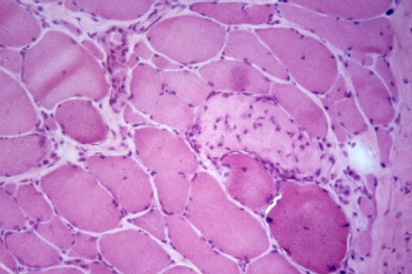
asmodium falciparum (80% of cases), Plasmodium vivax, Plasmodium ovale, Plasmodium malariae. Affects the red blood cells. Exists in tropical regions. Transmitted by the anopheles mosquito.

Rhinosporidiosis - (Latin name: Rhinosporidium seeberi). Affects the nose and nasopharynx. Exists in India and Sri Lanka. People who are bathing in common ponds can become contaminated when the nasal mucosa is in contact with infected material..

Toxoplasmosis. Parasitic pneumonia - (Latin name: Toxoplasma gondii). Affects the liver, heart, eyes and brain. Widespread worldwide. People can become infected after ingesting raw/undercooked pork, lamb, goat, milk. Also may exist in contaminated food or soil with cat feces.

Trichomoniasis - (Latin name: Trichomonas vaginalis). Affects the female urogenital tract. Can exist in males, but without symptoms. This is an STI (sexually transmitted infection).

Sleeping sickness - (Latin name: Trypanosoma brucei). Affects the central nervous system, blood lymph. Transmitted by the tsetse fly.

Chagas disease - (Latin name: Trypanosoma cruzi). Affects the blood, muscle, nerves, heart, esophagus and colon. Transmitted through an insect bite.

Helminths organisms (worms)

Ancylostomiasis/Hookworm - (Latin name: Ancylostoma duodenale, Necator americanus). Affects the lungs, small intestine, and blood.

Anisakiasis - (Latin name: Anisakis). The human has an allergic reaction. Human can become infected when eating contaminated fish and seafood.

Roundworm. Parasitic pneumonia - (Latin name: Ascaris sp. Ascaris lumbricoides). Affects the intestines, lungs, pancreas, liver, appendix (the patient develops Löffler's syndrome). Tropical and subtropical regions.

Roundworm - (Latin name: Baylisascariasis, Baylisascaris procyonis). Affects the brain, lungs, liver and intestines. Infection comes from contaminated raccon stools. North America (rare).

Tapeworm. Tapeworm infection - (Latin name: Cestoda). Affects the intestine. Today this parasite is rare.

Clonorchiasi - (Latin name: Clonorchis sinensis; Clonorchis viverrini, Dicrocoelium dendriticum). Affects the gall bladder. Humans can become infected after ingesting contaminated ants.

Dioctophyme renalis infection - (Latin name: Dioctophyme renale). Usually affects the right kidney. Exists all over the world. Humans can become infected after eating raw or undercooked freshwater fish.

Diphyllobothriasis. Tapeworm - (Latin name: Diphyllobothrium latum). Affects the intestines and blood. Exists in Chile, Peru, Japan, Uganda and Europe. Humans can become infected after eating raw freshwater fish.

Guinea worm. Dracunculiasis - (Latin name: Dracunculus medinensis). Person gets blisters and ulcers, affects subcutaneous tissues and muscle.

Echinococcosis .Tapeworm - (Latin name: Echinococcus granulosus, Echinococcus multilocularis, E. vogeli, E. oligarthrus). Affects the spleen, kidneys, lungs and liver. Mediterranean region. The human can become infected after eating foods contaminated by the feces of a carnivore, or eating offal from a herbivore.

Pinworm. Enterobiasis - (Latin name: Enterobius vermicularis, Enterobius gregorii). Affects the intestines and anus. This bacteria is more common in temperate regions.

Liver fluke. Fasciolosis - (Latin name: Fasciola hepatica, Fasciola gigantic). Affects the gall bladder and liver. Common in various parts of the world. Contamination comes from freshwater snails.

Fasciolopsiasis. Intestinal fluke - (Latin name: Fasciolopsis buski). Affects the intestines. Common in East Asia. When humans ingest infested water plants or water they can become infected.

Gnathostomiasis - (Latin name: Gnathostoma spinigerum, Gnathostoma hispidum). Affects people under the skin. It is rare, and occurs in Southeast Asia. Caused by eating freshwater fish, pigs, snails, frogs, and chicken.

Hymenolepiasis - (Latin name: Hymenolepis nana, Hymenolepis diminuta). Humans can become infected by ingesting material contaminated by cockroaches, meal worms and flour beetles.

Loa loa filariasis, Calabar swellings - (Latin name: Loa loa filarial). Affects the eyes, lungs and connective tissue. Mainly in West Africa. Transmitted by horsefly bites.

Mansonelliasis. Filariasis - (Latin name: Mansonella streptocerca). Affects the subcutaneous layer of the skin.

Metagonimiasis. Intestinal fluke - (Latin name: Metagonimus yokogawai. Caused by eating undercooked or salted fish. Found in Siberia, Balkan states, Israel, Spain and Manchuria.

River blindness - (Latin name: Onchocerca volvulus, Onchocerciasis). Affects the eyes, skin, and tissue. Found in Africa, Central/South America, near fast flowing, cool rivers. Transmitted by a fly.

Chinese Liver Fluke - (Latin name: Opisthorchis viverrini, Opisthorchis felineus, Clonorchis sinensis). Affects the bile duct. In Russia 1.5 million people are affected. Caused by eating raw, slightly salted or frozen fish.

Paragonimiasis, Lung Fluke - (Latin name: Paragonimus westermani; Paragonimus africanus; Paragonimus caliensis; Paragonimus kellicotti; Paragonimus skrjabini; Paragonimus uterobilateralis). Affects the lungs and sputum. Causd by eating undercooked or raw freshwater crabs, crayfishes and other crustaceans.

Schistosomiasis, bilharzia, bilharziosis or snail fever - (Latin name: Schistosoma sp.). Affects the skin. Caused by exposure to fresh water snail infested waters. Found in South America (eastern), Africa, the Caribbean, and the Middle East.

Intestinal schistosomiasis - (Latin name: Schistosoma mansoni). Affects the skin, spleen intestine, liver, and lungs. 83 million people are affected. Found in the Middle East, Asia, South America, Africa and the Caribbean. Human skin exposed to water infested with a type of fresh water snail can become infected.

Urinary schistosomiasis - (Latin name: Schistosoma haematobium). Affects the skin, lungs, ureters, bladder and kidneys. Found in the Middle East and Africa. Humans can become infected if their skin is exposed to water contaminated with a type of snail.

Schistosomiasis by Schistosoma japonicum - (Latin name: Schistosoma japonicum). Affects the intestine, skin, spleen, and liver. Humans can become infected if their skin is exposed to water contaminated with a type of snail.

Asian intestinal schistosomiasis - (Latin name: Schistosoma mekongi). Found in southeast Asia. Humans can become infected if their skin is exposed to water contaminated with a type of snail.

Sparganosis - (Latin name: Spirometra erinaceieuropaei). Humans can become infected if they eat foods tainted with dog or cat feces.

Strongyloidiasis.Parasitic pneumonia - (Latin name: Strongyloides stercoralis). Affects the lungs, skin and intestines. The parasite penetrates through the skin.

Beef tapeworm - (Latin name: Taenia saginata). Exists all over the world. Affects the intestines. Caused by eating undercooked beef.

Pork tapeworm - (Latin name: Taenia solium). Caused by eating undercooked pork.

Toxocariasis - (Latin name: Toxocara canis, Toxocara cati). Affects the eyes, brain and liver. Caused by eating Toxocara egg tainted foods.

Trichinosis - (Latin name: Trichinella spiralis, Trichinella britovi, Trichinella nelsoni, Trichinella native). Affects muscle, small intestine and periobital region. Caused by eating undercooked pork.

Swimmer's itch - (Latin name: Trichobilharzia regenti, Schistosomatidae). Affects the skin. Caused by going into contaminated water.

Whipworm - (Latin name: Trichuris trichiura, Trichuris vulpis). Affects the large intestine and anus. It is common all over the world. Humans can become infected when ingesting the eggs in dry goods, for example rice, several grains, beans, or soil tainted with human excrements.

Elephantiasis Lymphatic filariasis - (Latin name: Wuchereria bancrofti, lymphatic system). Caused by mosquito bites.

Ectoparasites

Bedbug - (Latin name: Cimicidae Cimex lectularius). Affects the skin and vision. Found all over the world. Sharing clothing and bedding can spread infection.

Head louse. Pediculosis - (Latin name: Pediculus humanus). Affects the hair follicles. Common all over the world. Spread by head-to-head contact.

Body louse. Pediculosis - (Latin name: Pediculus humanus corporis). Common all over the world. Infection can spread by sexual activity, skin-to-skin contact, and sharing bedding and/or clothing.

Crab louse. Pediculosis - (Latin name: Phthirus pubis). Affects the pubic area and eyelashes. Common all over the world. Infection can spread by sexual activity, skin-to-skin contact, and sharing bedding and/or clothing.

Demodex. Demodicosis - (Latin name: Demodex folliculorum/brevis/canis). Affects the eyebrow and eyelashes. Common all over the world. Caused by prolonged skin contact.

Scabies - (Latin name: Sarcoptes scabiei). Affects the skin. Common all over the world. Infection can spread by sexual activity, skin-to-skin contact, and sharing bedding and/or clothing.

Screwworm. Cochliomyia - (Latin name: Cochliomyia hominivorax). Affects skin and wounds. Found in Central America and north Africa. Transmitted by a fly.

Wheat Allergy

People with a wheat allergy have an abnormal immune system response to at least one of the proteins that exist in wheat. It is one of the most common childhood food allergies, but may affect adults as well. The person with a wheat allergy has developed a specific antibody to a wheat protein, and sometimes more than one.

People with wheat allergies can respond with a variety of possible signs and symptoms, including breathing difficulties, nausea, hives, bloated stomach and an inability to focus. With some people the consumption of wheat and wheat products may result in anaphylaxis - a life-threatening allergic response.

The allergic reaction involves IgE (immunoglobulin) antibodies to at least one of the following proteins found in wheat:
  • Albumin
  • Globulin
  • Gliadin
  • Glutenin (gluten)
Most allergic reactions involve albumin and globulin. Allergy to gliadin and gluten are less common. Gluten allergy is often confused with Celiac disease or some other digestive disorders.

Some people have an allergic reaction when they inhale wheat flour, while others need to eat it in order to experience symptoms. An allergic reaction can occur within minutes or sometimes hours of either consuming or inhaling wheat.

What are the signs and symptoms of wheat allergy?

A symptom is something the patient feels and reports, while a sign is something other people, such as the doctor detect. For example, pain may be a symptom while a rash may be a sign. The most common signs and symptoms of a wheat allergy include:
  • Allergic rhinitis - nasal congestion
  • Anaphylaxis
  • Asthma
  • Atopic dermatitis (eczema)
  • Diarrhea
  • Mouth irritation and possible swelling
  • Nausea
  • Throat irritation and possible swelling
  • Urticaria - hives, an itchy rash (possible swelling of the skin)
  • Vomiting
  • Watery and/or itchy eyes
  • Bloated stomach
Signs and symptoms of anaphylaxis may include:
  • Throat swells and becomes tight
  • Chest tightness
  • Chest pain
  • More severe breathing difficulties
  • Swallowing difficulties
  • Skin becomes pale or bluish
  • Weak pulse
  • Hypotension - a serious drop in blood pressure
Anaphylaxis is a medical emergency.

Vitamin B12 Deficiency

What Is Vitamin B12 Deficiency? What Causes Vitamin B12 Deficiency


Vitamin B12 is crucial for the proper formation of red blood cells, as well as the health of nerve tissue. Vitamin B12 deficiency, or B12 deficiency, if left untreated can result in anemia, as well as irreversible nerve and brain damage.

A lack of vitamin B12 in the blood can lead to a blood disorder called pernicious anemia. Those with the disorder are unable to produce enough of a protein substance - IF (intrinsic factor) - in their stomach that allows their body to absorb vitamin B12. Vitamin B12 is injected straight into the patient's blood, therefore bypassing the stomach which is unable to absorb it properly.

Apart from creating red blood cells and keeping our nervous system healthy, we also need vitamin B12 in order to be able to absorb folic acid. Vitamin B12 also helps to release energy.

According to Medilexicon's medical dictionary:
    Vitamin B12 is a "generic descriptor for compounds exhibiting the biologic activity of cyanocobalamin; the antianemia factor of liver extract that contains cobalt, a cyano group, and corrin in a cobamide structure. Several substances with similar formulas and with the characteristic hematinic action have been isolated and designated: B12a, hydroxocobalamin; B12b, aquacobalamin; B12c, nitritocobalamin; B12r, cob(II)alamin; B12s, cob(I)alamin; B12III, factors A and V1a (cobyric acid) and pseudovitamin B12. Vitamins B12a and B12b are known to be tautomeric compounds; B12b has been obtained from cultures of Streptomyces aureofaciens; B12c has been obtained from cultures of Streptomyces griseus and is distinguishable from B12 by differences in its absorption spectrum. The physiologically active vitamin B12 coenzymes are methylcobalamin and deoxyadenosinecobalamin. A deficiency of vitamin B12 is often associated with certain methylmalonic acidurias."

What are the signs and symptoms of vitamin B12 deficiency?

A symptom is something the patient feels and reports, while a sign is something other people, such as the doctor detect. For example, pain may be a symptom while a rash may be a sign.

Vitamin deficiencies tend to develop gradually and slowly; sometimes over a period of several years. At first, signs and symptoms may be subtle - but as time goes by they become more noticeable.

Our cells need vitamin B12 in order to multiply properly. We produce millions of red blood cells every minute. A vitamin B12 deficiency affects the production of red blood cells. Subsequently, the red blood cell count drops and the patient develops anemia. The most common symptoms of anemia are:
  • Fatigue (tiredness)
  • Panting (shortness of breath)
  • Palpitations (disagreeable sensations of irregular and/or heavy beating of the heart)
B12 deficiency anemia may also be caused by a lack of intrinsic factor - pernicious anemia. The patient's digestive system cannot absorb B12 properly. Signs and symptoms of pernicious anemia may include:
  • Fatigue
  • Shortness of breath
  • Palpitations
Anemia may also have the following signs and symptoms:
  • A sore mouth and/or tongue
  • Weight loss
  • Pale or yellowish skin
  • Diarrhea (sporadic)
  • Menstrual problems
  • Higher susceptibility to infections
If the deficiency continues untreated the patient may have the following neurological signs and symptoms:
  • Tingling or numbness of the fingers
  • Tingling or numbness of the toes
  • General muscle weakness
  • Difficulty walking properly (staggering)
  • Irritability
  • Confusion
  • Forgetfulness
  • Tender calves

What are the causes of vitamin B12 deficiency?

Causes of pernicious anemia - pernicious anemia is caused by an autoimmune disease; the person's own immune system attacks good parts of the body, as if they were bacteria or viruses.
  • The immune system of patients with pernicious anemia creates antibodies which attack the lining of the stomach, damaging cells that produce intrinsic factor. Intrinsic factor is a substance that is secreted by the gastric mucous membrane (lining of the stomach) and is vital for the absorption of vitamin B12 in the intestines. If the production of intrinsic factor is undermined, vitamin B12 cannot be absorbed into the body properly.

  • Bowel problem - patients with Crohn's disease, as well as people who have had their bowels surgically shortened, may have problems absorbing vitamin B12 into their bloodstream. Short bowel syndrome is a term used for people who have had their bowel shortened (half or more of their small intestine removed). Patients with short bowel syndrome typically experience diarrhea, cramping and heartburn. It is not uncommon for individuals to become malnourished because what is left of the small intestine cannot absorb adequate quantities of vitamins, water and other nutrients.

  • Vegan diet - some people who follow a vegan diet may suffer from B12 deficiency if they do not eat fortified foods, or some types of yeast.

How is B12 deficiency diagnosed?

A GP (general practitioner, primary care physician) will interview the patient and carry out a physical examination, looking out for such signs as an accelerated pulse and pale/yellowish skin.
  • Blood test - to determine whether the red blood cell count is low. The appearance of the red blood cells is also checked, because they may have an unusual shape. A blood test can also determine what the level of B12 is.

    The GP may also want to check the patient's blood intrinsic factor antibody levels, in order to determine whether the person has pernicious anemia.

  • Bone marrow biopsy - the aim here is to rule out other possible causes of red cell abnormalities or anemia.

What are the treatment options for B12 deficiency?

Hydroxocobalamin injections - this is a form of B12. It is injected into a muscle every two to four days. After about six injections the patient should have a good store of B12.

In the majority of cases the patient will notice significant improvements in symptoms soon after receiving injections.

Annual blood tests are common, to monitor the success of treatment.

Most patients require booster injections of B12 every three months for the rest of their lives.

Prevention of B12 deficiency

  • Vegans and some vegetarians - vegetarians who do not eat eggs, as well as vegans should make sure their B12 intake is adequate. There are various breakfast cereals which are fortified with vitamin B12. Some brands of nutritional yeast are also good sources of B12. Some types of soy milk are fortified with B12.

  • People who eat meat and/or fish - a balanced diet containing fish, meat and dairy foods should have enough B12 for human requirements.

Hepatitis


  1. What Is Hepatitis? Symptoms, Causes and Treatments


The word hepatitis comes from the Ancient Greek word hepar (root word hepat) meaning 'liver', and the Latin itis meaning inflammation. Hepatitis means injury to the liver with inflammation of the liver cells.

What is the liver?

The liver is the largest gland in the human body. It weighs approximately 3 lb (1.36 kg). It is reddish brown in color and is divided into four lobes of different sizes and lengths. It is also the largest internal organ (the largest organ is the skin). It is below the diaphragm on the right in the thoracic region of the abdomen. Blood reaches the liver through the hepatic artery and the portal vein. The portal vein carries blood containing digested food from the small intestine, while the hepatic artery carries oxygen-rich blood from the aorta.

The liver is made up of thousands of lobules, each lobule consists of many hepatic cells - hepatic cells are the basic metabolic cells of the liver.

The liver has a wide range of functions, including:
  • Detoxification (filters har mful substances form the blood, such as alcohol)
  • Stores vitamins A, D, K and B12 (also stores minerals)
  • Protein synthesis (makes certain amino acids - the building blocks of proteins)
  • The production of biochemicals needed for digestion, such as bile
  • Maintains proper levels of glucose in the blood
  • Produces 80% of your body's cholesterol (cholesterol is vital)
  • The storage glycogen (also converts glucose to glycogen)
  • Decomposing red blood cells
  • Synthesizing plasma protein
  • The production of hormones
  • Produces urea (the main substance of urine)
Hepatitis can heal on its ow n with no significant consequence, or it can progress to scarring of the liver. Acute hepatitis lasts under six months, while chronic hepatitis lasts longer.

Most liver damage is caused by 3 hepatitis viruses, called hepatitis A, B and C. However, hepatitis can also be caused by alcohol and some other toxins and infections, as well as from our own autoimmune process (the body attacks itself).

About 250 million people globally are thought to be affec ted by hepatitis C, while 300 million people are thought to be carriers of hepatitis B.

Not all forms of hepatitis are infectious. Alcohol, medicines, and chemical may be bad for the liver and cause inflammation. A person may have a genetic problem, a metabolic disorder, or an immune related injury. Obesity can be a cause of liver damage which can lead to inflammation. These are known as non-infectious, because they cannot spread form person-to-person.

How many types of hepatitis are there?

There are five main types of hepatitis that are caused by a virus, A, B, C, D, and E - plus types X and G.
  • Hepatitis A - this is caused by eating infected food or water. The food or water is infected with a virus called HAV (Hepatitis A Virus). Anal-oral contact during sex can also be a cause. Nearly everyone who develops Hepatitis A makes a full recovery - it does not lead to chronic disease.


  • Hepatitis B - this is an STD (sexually transmitted disease). It is caused by the virus HBV (Hepatitis B Virus) and is spread by contact with infected blood, semen, and some other body fluids. You get Hepatitis B by:
    • Unprotected sexual intercourse with an infected person (unprotected sex means without using a condom) Using a syringe that was previously used by an infected person (most commonly happens with drug addicts and people who inject steroids).

    • Having your skin perforated with unsterilized needles, as might be the case when getting a tattoo, or being accidentally pricked. People who work in health care risk becoming infected by accident in this way. Sharing personal items, such as a toothbrush or razor, with an infected person.

    • A baby can become infected through his mother's milk if she is infected.

    • Being bitten by someone who is infected.

    The liver of a person infected with Hepatitis B swells. The patient can suffer serious liver damage due to infection, resulting in cancer. For some patients the hepatitis becomes chronic (very long-term or lifelong). Donated blood is always tested for Hepatitis B.
  • Hepatitis C - Hepatitis C is usually spread through direct contact with the blood of a person who has the disease. It is caused by the virus HCV (Hepatitis C Virus). The liver can swell and become damaged. In hepatitis C, unlike hepatitis B, liver cancer risk is only increased in people with cirrhosis and only 20% of hep C patients get cirrhosis. Feces is never a route of transmission in hepatitis C. Donated blood is also tested for Hepatitis C.
  • Hepatitis D - only a person who is already infected with Hepatitis B can become infected with Hepatitis D. It is caused by the virus HDV (Hepatitis D Virus). Infection is through contact with infected blood, unprotected sex, and perforation of the skin with infected needles. The liver of a person with Hepatitis D swells.
  • Hepatitis E - a person can become infected by drinking water that contains HEV (Hepatitis E Virus). The liver swells but there is no long-term consequence. Infection is also possible through anal-oral sex.

  • Hepatitis X - if a hepatitis cannot be attributed to the viruses of hepatitis A, B, C, D, or E, it is called Hepatitis X. In other words, hepatitis of an unknown virus.
  • Hepatitis G - this is a type of hepatitis caused by the Hepatitis G virus (HGV). Usually there are no symptoms. When there are symptoms they are very mild.

What are the signs and symptoms of hepatitis?

Many people with Hepatitis experience either mild symptoms or none at all. Remember that an infected person's feces are always infectious to other people. When symptoms appear, they usually do so about 15 to 180 days after the person has become infected.

The acute phase of hepatitis - symptoms

The initial phase of hepatitis is called the acute phase. The symptoms are like a mild flu, and may include:
  • Diarrhea
  • Fatigue
  • Loss of appetite
  • Mild fever
  • Muscle or joint aches
  • Nausea
  • Slight abdominal pain
  • Vomiting
  • Weight loss
The acute phase is not usually dangerous, unless it develops into the fulminant or rapidly progressing form, which can lead to death.

As the patient gets worse, these symptoms may follow:
  • Circulation problems (only toxic/drug-induced hepatitis)
  • Dark urine
  • Dizziness (only toxic/drug-induced hepatitis)
  • Drowsiness (only toxic/drug-induced hepatitis)
  • Enlarged spleen (only alcoholic hepatitis)
  • Headache (only toxic/drug-induced hepatitis)
  • Hives
  • Itchy skin
  • Light colored feces, the feces may contain pus
  • Yellow skin, whites of eyes, tongue (jaundice)
Patient outcomes after the acute phase depend on various factors, especially the type of hepatitis.

Treatments for hepatitis

  • Hepatitis A - There is no treatment specifically for hepatitis A. Doctor will advise the patient to abstain from alcohol and drugs during the recovery. The vast majority of patients with Hepatitis A will recover spontaneously.

  • Hepatitis B - A patient with Hepatitis B needs to rest. He will require a diet that is high in protein and carbohydrate - this is to repair damaged liver cells, as well as to protect the liver. If this is not enough, the doctor may prescribe interferon. Interferon is an antiviral agent.

  • Hepatitis C - A patient with Hepatitis C will be prescribed pegylated interferon and ribavirin.

  • Hepatitis D or E - So far, there is no effective treatment for either Hepatitis D or E.

  • Non-Viral Hepatitis - If the patient has non-viral hepatitis, the doctor needs to remove the harmful substance. It will be flushed out of the stomach by hyperventilation or induced vomiting. Patients with drug-induced hepatitis may be prescribed corticosteroids.

How to prevent hepatitis

How to prevent Hepatitis A
  • Wash your hands with soap after going to the toilet
  • Only consume food that has just been cooked
  • Only drink commercially bottled water, or boiled water if you unsure of local sanitation
  • Only eat fruits that you can peel if you are somewhere where sanitation is unreliable
  • Only eat raw vegetables if you are sure they have been cleaned/disinfected thoroughly
  • Get a vaccine for Hepatitis A if you travel to places where hepatitis may be endemic

How to prevent Hepatitis B
  • Tell the partner if you are a carrier or try to find out whether he/she is a carrier
  • Practice safe sex
  • Only use clean syringes that have not been used by anyone else
  • Do not share toothbrushes, razors, or manicure instruments
  • Have a Hepatitis B series of shots if you are at risk
  • Only allow well sterilized skin perforating equipment (tattoo, acupuncture, etc.)

How to prevent Hepatitis C
  • If you are infected do not let others share your toothbrush, razor, manicure equipment
  • If you are infected cover open wounds
  • Do not share needles, toothbrushes, or manicure equipment
  • If your skin is to be pierced, make sure equipment is well sterilized (tattoo, etc.)
  • Go easy on the alcohol
  • Do not share drug equipment

How to prevent Hepatitis D
  • Use the same guidelines as for Hepatitis B. Only a person who is infected with Hepatitis B can become infected with Hepatitis D.
How to prevent Hepatitis E
  • Do the same as you would to protect yourself from Hepatitis A infection.
How to prevent Alcoholic HepatitisLink
  • Go easy on the alcohol, or abstain from consuming alcohol
How to prevent Toxic/Drug Induced Hepatitis
  • Make sure you know about the lethal contents of all chemicals
  • Make sure the spray is not pointing at you
  • Make sure you wear protective gear if you have to
Share |